Quantitative Liver Imaging
Pulse-echo ultrasound-based liver fat fraction assessment based on quantitative sound speed estimation and aberration correction
Early Liver Sound Speed Estimation Based on Layered Abdomen Model


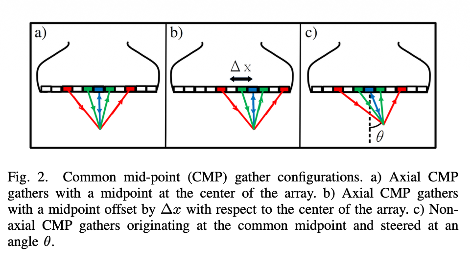
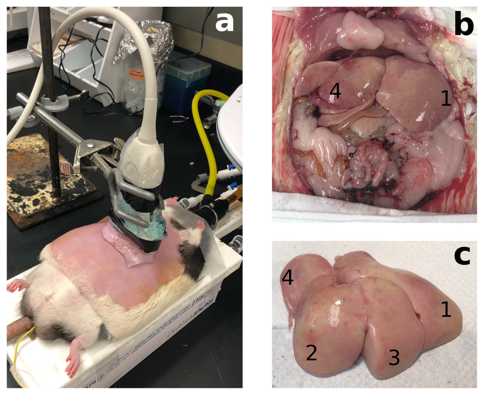
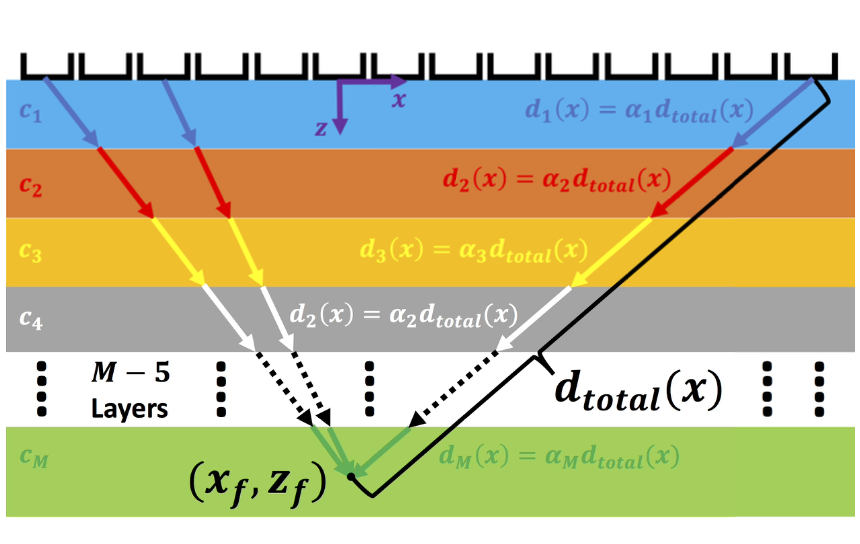
Early work focused on estimating liver sound speed from pulse-echo ultrasound using a layered model of the abdomen (Ali et al., 2021; Ali et al., 2020; Ali et al., 2020; Brevett et al., 2022). The beamforming sound speed that produced the best focus at a given depth was related to the average sound speed of the tissue above that depth, allowing the measured depth-dependent values to be inverted to estimate a local sound-speed profile. This approach was demonstrated in obese Zucker rats with different grades of hepatic steatosis (Ali et al., 2021; Telichko et al., 2022). The estimated liver sound speeds agreed closely with measurements from the corresponding excised liver samples, demonstrating the potential of pulse-echo ultrasound for quantitative assessment of liver tissue.


Quantitative Full-Wave Estimation of Liver Sound Speed


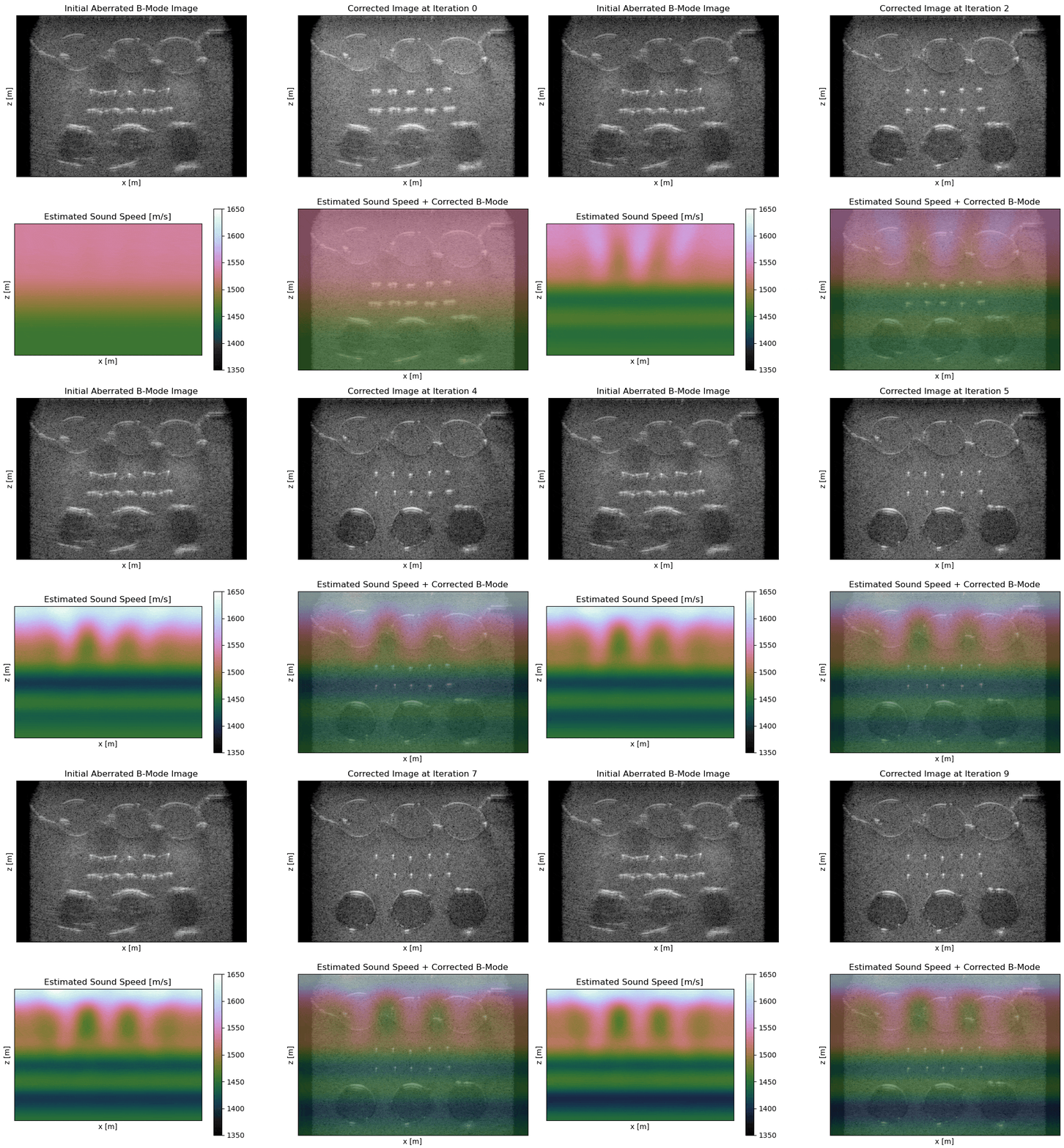
The layered-medium model provides a useful estimate of liver sound speed, but it cannot adequately describe the lateral sound-speed variations encountered in the abdomen. These variations produce both travel-time errors and diffractive effects that degrade the image when conventional beamforming assumes a constant sound speed. To address this limitation, I developed wave-equation migration velocity analysis (WEMVA) for sound-speed estimation and aberration correction in pulse-echo ultrasound. WEMVA uses reverse-time migration (RTM) to reconstruct the image by cross-correlating the transmitted and backpropagated received wavefields. Because the RTM image is differentiable with respect to the sound-speed distribution, the image-domain error can be used to iteratively update the sound speed estimate.
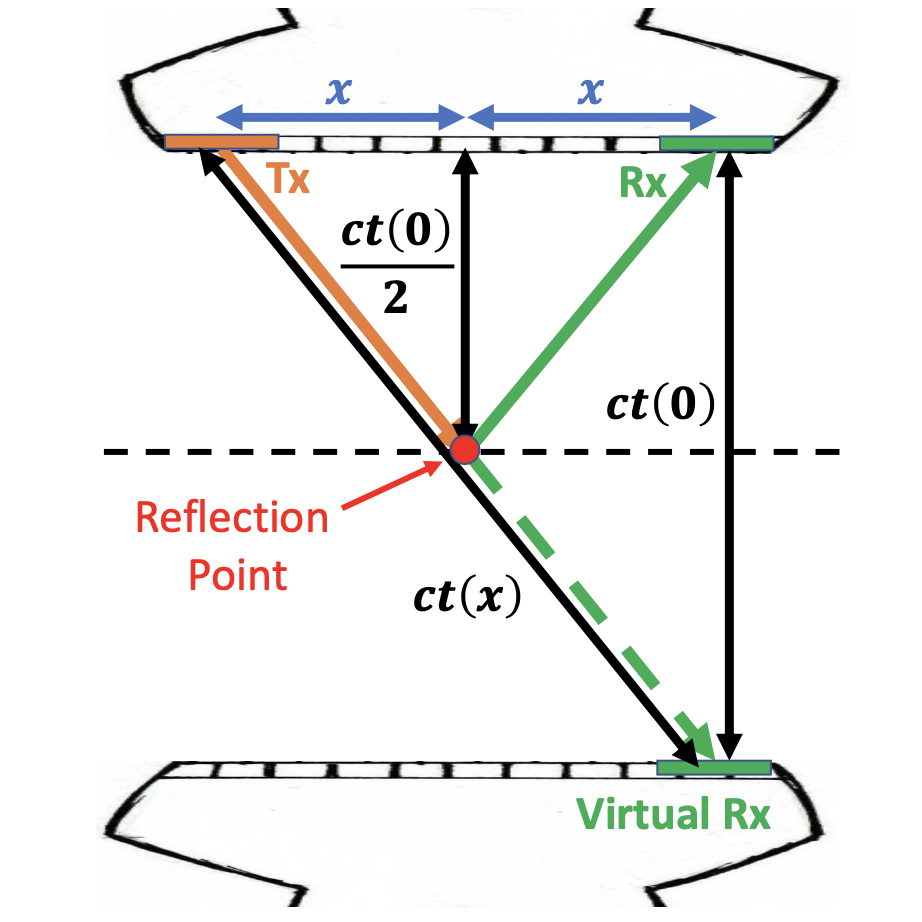
My most recent form of WEMVA uses extended RTM images with a subsurface offset between the transmit and receive wavefields. The correct sound speed profile should focus the extended image at zero subsurface offset; sound speed errors produce residual energy at nonzero subsurface offsets. Driving this energy toward zero subsurface offset provides an image-domain criterion for estimating the sound speed profile (Ali et al., 2026).


Future Extension of WEMVA to Curvilinear Human-Abdominal Imaging
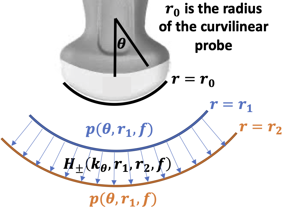
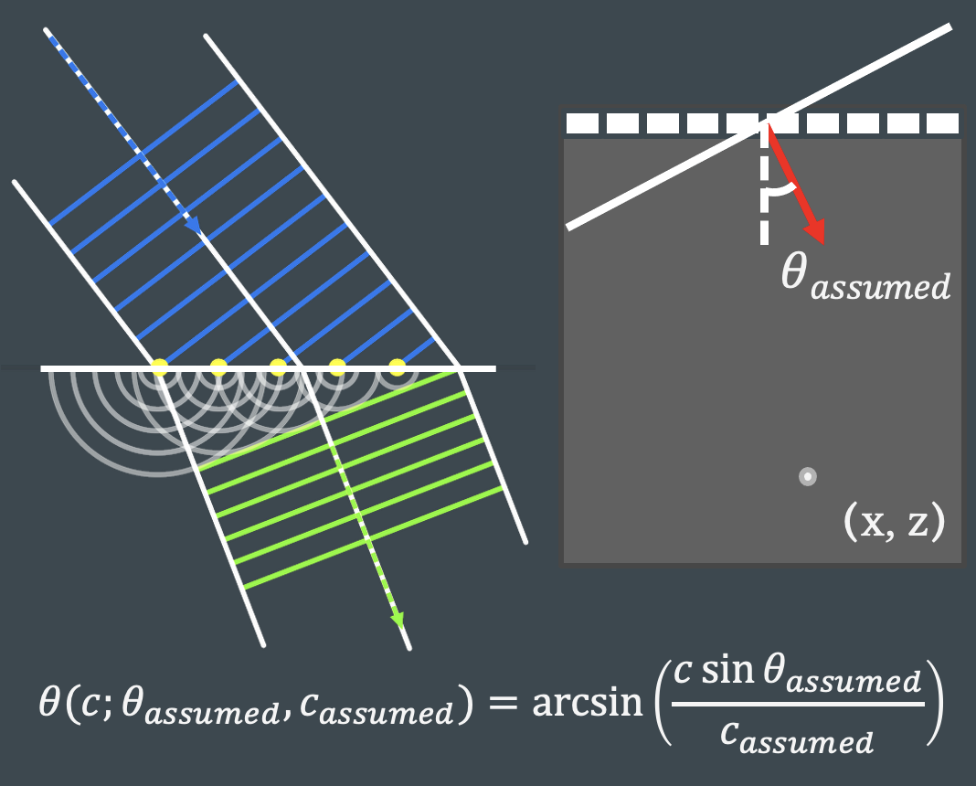
The initial WEMVA formulation was developed for linear arrays, whereas clinical abdominal ultrasound commonly uses curvilinear probes. Extending WEMVA to these probes requires the wave-propagation model to account for the curved transducer geometry rather than treating the aperture as planar. I have previously extended the angular spectrum method used in RTM to a polar coordinate system for curvilinear arrays (Ali & Dahl, 2022). The formulation propagates the transmitted and received wavefields in the polar geometry of the curved probe, while retaining the Fourier-domain efficiency of the angular spectrum method. This provides the wave-propagation engine needed to apply RTM and WEMVA directly to curvilinear abdominal acquisitions. The underlying curvilinear angular-spectrum formulation was validated against Field II simulations and demonstrated using in-vivo abdominal channel data. Combining this propagation model with WEMVA would enable sound speed estimation and aberration correction using conventional curvilinear abdominal imaging probes.